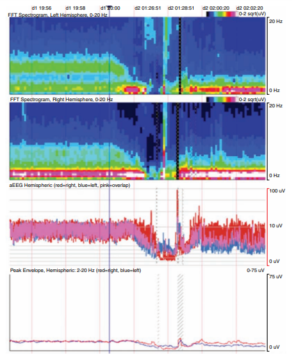
The following quantitative EEG (qEEG) fast Fourier transform (FFT) spectrograms show 0–20 Hz activity from the left and right hemispheres (top 2 panels), amplitude-integrated EEG, and peak envelope; the color bar indicates the power, black being the lowest and white being the highest. Note that the red vertical lines represent 2-minute intervals; for illustrative purposes, 3 different time periods are spliced together. What is the best explanation of what occurred after the blue vertical line indicator?
A. The patient had a seizure with postictal slowing afterwards
B. The patient had diffuse cerebral ischemia due to cardiac arrest with subsequent infarction
C. The patient was disconnected from continuous video-EEG (cEEG) monitoring temporarily to be taken to CT scan without any significant interim clinical event
D. The patient transitioned from a wakeful state to deeper stages of normal sleep