PY162 PAIN CASE LABORATORY EXERCISE |
Demonstration to investigate some sites of drug action on gastrointestinal tract motility. |
Objectives
- Understand the requirements for maintaining the functioning of ex vivo tissue.
- Understand the pharmacological regulation of G I motility
- Understand the pharmacological and physiological regulation of autonomic control of the gut.
- Appreciate the rationale for the uses of drugs that regulate gut function in the treatment of disease.
Introduction
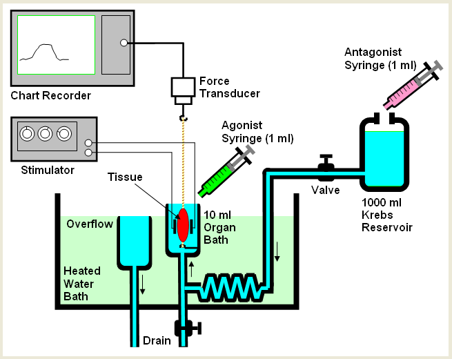
Pharmacists are the experts in the healthcare team on medicines and it is essential that they understand the how drug molecules affect the function of different organ systems in the body. The aim of this demonstration is to illustrate the functional role of different drugs on gastrointestinal tract motility using a section of guinea pig colon. You will examine the effects of a variety of drugs e.g. morphine or naloxone on the contractions of the guinea pig ileum evoked by trans-mural stimulation. Sections of the gastrointestinal tract can be stimulated by passing a current across the muscular wall of the tissue (trans-mural stimulation). A diagram of the structure of the gastrointestinal tract is shown below.
As you can see from the diagram the gastrointestinal tract is a complicated piece of tissue containing two layers of muscle and a number of different plexii that contain nerve cells which help to regulate muscle contractions. A photomicrograph of part of a plexus is shown below.
Providing the properties of the current pulse are precisely controlled it is possible to selectively stimulate the nerve cells to release their neurotransmitter and drive the muscle to contract.
In the second part of the practical you will observe how morphine affects the motility of an artificial faecal pellet in an ex vivo preparation of the intact guinea pig colon.
Method
1. Analysis of the effects of a range of drug molecules on evoked contractions of the guinea pig ileum.
- Prepare a piece of guinea pig ileum for transmural stimulation. (see diagram below)
- Starting with a pulse width of 0.5ms, frequency 0.1Hz, gradually increase the voltage until you obtain a measurable contraction.
- Add the morphine (10mM stock) or atropine (10mM stock) to the bath (final concentration 1mM).
- With the morphine or atropine still in the bath increase the pulse width on the stimulator until you record a contraction. Under these circumstances you are selectively stimulating the muscle. B. The muscle has no voltage-gated Na+ channels.
- Apply morphine in the presence of naloxone or atropine and observe the effects on muscle contraction.
- Apply KCl (50mM bath concentration)
- Effects of various drugs on colonic motility
- A freshly dissected piece of guinea pig colon is placed in a long organ bath. Small incisions are made in both ends of the colon and the tissue pinned to the sylgard-lined bath. These incisions will facilitate pellet insertion and evacuation from the colon.
- The preparation is continually perfused with warmed Krebs buffer solution.
- An epoxy-coated pellet is inserted into the proximal end of the colon using a blunt glass probe.
- Pellet motility through the bowel is recorded using a stop watch.
- 1 µM morphine is added to the perfusate and the effects on pellet motility determined.
- The effects of further adding 1 µM naloxone will be examined.
Important information
To ensure that you can complete the report successfully you should record the following information from the class:
- Ensure you have details of all the equipment used in the laboratory
- Describe and explain the effect of field stimulation on ileum contraction.
- Explain the effects of morphine on field stimulated contractions and how/why is this affected by naloxone.
- Explain how morphine affects the rate of pellet motility in the colon and how this is altered by the application of naloxone.
- Draw the following graphs from the experiments and provide detailed figure legends
- Molar concentration morphine versus contractile response of field stimulated tissue (in mN).
- The logarithm of the molar concentration morphine versus contractile response of field stimulated tissue (in mN).
- Molar concentration morphine versus % relaxation of stimulated tissue.
- The logarithm of the molar concentration morphine versus % relaxation of field stimulated tissue (in mN).
- Graph of pellet motility in the presence of morphine and following the application of naloxone.
Graphs must be generated using graphing software with responses in mN plotted against the Log10 of the concentration of the agonist.
What We Offer:
• On-time delivery guarantee
• PhD-level professionals
• Automatic plagiarism check
• 100% money-back guarantee
• 100% Privacy and Confidentiality
• High Quality custom-written papers


